
When your dentist recommends dental X-rays, you may wonder whether they are truly necessary, especially when your teeth feel fine and nothing appears wrong. While not every patient needs X-rays at every appointment, these images can reveal dental problems that cannot be seen during a visual examination alone. Dental X-rays allow your dentist to evaluate areas between the teeth, beneath existing restorations, around the roots, and within the jawbone. They may reveal early tooth decay, infections, bone loss, impacted teeth, and other concerns before they cause noticeable pain or visible damage.
At W Plus Dental, dental X-rays are recommended based on your individual oral health, symptoms, history, and treatment needs. The goal is not to take unnecessary images. It is to gather the information needed to provide an accurate diagnosis and help protect your long-term oral health.
Dental X-rays, also called dental radiographs, are diagnostic images of your teeth, roots, jawbone, and surrounding oral structures. They provide information that cannot always be obtained through a standard visual examination. The American Dental Association explains that dental X-rays can help identify damage or disease that may not be visible during a clinical dental exam, including new cavities and impacted teeth.
Dental X-rays are often used alongside a comprehensive dental checkup and cleaning to give your dentist a more complete understanding of your oral health.
No. You may not need dental X-rays at every appointment. Current American Dental Association recommendations emphasize that dental X-rays should be taken only when clinically necessary. Before recommending imaging, your dentist will consider your medical and dental history, previous X-rays, current symptoms, risk factors, and findings from your clinical examination.
Some patients may need X-rays more frequently because they have active decay, gum disease, extensive dental work, dental pain, or a higher risk of developing new problems. Patients with consistently healthy teeth and gums may be able to go longer between images.
Your recommended schedule may depend on factors such as:
There is no single X-ray schedule that is appropriate for every patient.
A visual dental exam provides valuable information, but your dentist cannot see through the enamel, beneath the gums, or inside the jawbone without diagnostic imaging.
Decay can develop between neighboring teeth where it may not be visible during a routine examination. Bitewing X-rays can help identify these cavities while they are still relatively small.
Early cavity detection may allow the tooth to be treated with a conservative tooth-colored dental filling. If decay progresses and weakens a larger portion of the tooth, a dental crown or another restorative treatment may become necessary.
An existing restoration may look normal from the outside while decay develops around its edges or underneath it. X-rays can help your dentist evaluate the tooth structure supporting a filling or crown.
A tooth infection may begin inside the tooth and spread into the surrounding bone. Dental X-rays can reveal changes around the root that may indicate an abscess or the need for root canal therapy.
Gum disease affects more than the visible gum tissue. Advanced periodontal disease can damage the bone that supports the teeth. X-rays help your dentist evaluate bone levels and determine whether you may benefit from periodontal treatment or a deeper dental cleaning.
Panoramic X-rays can show teeth that have not fully erupted, including impacted wisdom teeth. They also provide a broader view of the jaws and surrounding structures.
Some dental injuries are difficult to evaluate visually. Depending on the location and type of damage, imaging may help your dentist assess the tooth root, surrounding bone, or signs of infection.
For sudden pain, swelling, or a damaged tooth, contact us, an emergency dentist in Washington, DC.
Dental implants must be carefully planned around the available bone and nearby anatomical structures. In certain cases, three-dimensional cone-beam computed tomography, or CBCT imaging, may be used to evaluate the jaw and plan implant placement.
CBCT provides three-dimensional information, but it typically involves more radiation than conventional dental X-rays. For that reason, the FDA and ADA recommend using it only when the necessary diagnostic information cannot be obtained through lower-exposure imaging.
Learn more about dental implants in Washington, DC.
The type of image your dentist recommends will depend on the area being evaluated and the clinical question that needs to be answered.
Bitewing X-rays show the upper and lower back teeth in a specific section of the mouth. They are commonly used to check for cavities between teeth and evaluate the bone level around them.
A periapical X-ray shows the entire tooth, including the crown, root, and surrounding bone. It may be recommended when evaluating dental pain, trauma, infection, or a specific tooth.
A panoramic image provides a broad view of the upper and lower jaws, teeth, sinuses, and surrounding structures. It may be used to assess wisdom teeth, missing teeth, jaw conditions, or general dental development.
CBCT creates a detailed three-dimensional image. It may be recommended for dental implant planning, complex root canal treatment, impacted teeth, or other situations that require more detailed anatomical information.
Dental X-rays use very low levels of radiation. Modern equipment, digital imaging, focused beam sizes, and proper patient positioning help keep exposure as low as reasonably achievable.
The ADA states that radiation doses from dental imaging are much lower than they were in the past because of advances in technology. The organization still recommends using X-rays in moderation and taking them only when they provide necessary diagnostic information. The small radiation risk should always be considered alongside the potential benefit of detecting a cavity, infection, bone defect, or other condition that may otherwise remain untreated.
Digital dental X-rays offer several benefits for both patients and dentists. The images are available almost immediately and can be enlarged or adjusted to help evaluate specific areas more closely.
Digital images can also be stored in your dental record, compared with future images, and transferred between dental offices when necessary. The ADA recommends making an effort to use previous dental images when they are still current and diagnostically useful, helping prevent unnecessary repeat exposure.
When visiting a new dentist, ask your previous dental office to send recent X-rays. Your new dentist can review them and decide whether additional images are actually needed.
Even when you are not experiencing pain, new-patient X-rays may be recommended to establish a clear picture of your current oral health.
Your dentist may use these images to:
However, recent images from your previous dentist may provide the information needed. Bring or transfer your records before your first dental exam in DC whenever possible.
You always have the right to ask questions and discuss your concerns before agreeing to dental X-rays. However, declining recommended imaging may limit your dentist’s ability to diagnose certain conditions. A cavity between the teeth, infection around a root, or bone loss from gum disease may not be visible without an X-ray.
We will explain:
The ADA encourages dentists and patients to discuss the need for X-rays and make informed treatment decisions together.
Preventive dental care is about identifying concerns before they become more painful, complicated, or expensive to treat. Dental X-rays can be an important part of that process when they are combined with a thorough examination, professional cleaning, and personalized treatment recommendations.
For example, a small cavity found between the teeth may be treated with a filling. Without imaging, that same cavity could continue growing until the tooth requires a crown, root canal, or extraction.
Dental X-rays are not a replacement for an in-person examination. They are one component of a complete diagnostic process.
There is no universal schedule for every adult. The frequency depends on your oral health, cavity risk, gum health, symptoms, treatment history, and the availability of recent diagnostic images. Patients with active dental problems may need imaging more frequently than patients with consistently healthy teeth and gums.
Yes. Dental X-rays can reveal cavities between teeth or beneath restorations before they cause pain. Tooth decay often produces few symptoms during its earliest stages.
X-rays can show changes in the bone supporting the teeth. This information helps your dentist evaluate the severity and progression of periodontal disease when combined with gum measurements and a clinical examination.
According to the ADA’s patient guidance, necessary dental X-rays can generally be taken during pregnancy because they use very low doses of radiation and the beam is focused on the area being examined. Tell your dental team when you are pregnant or think you may be pregnant so your care can be appropriately planned.
Current ADA recommendations state that routine abdominal and thyroid shielding is no longer necessary with modern dental X-ray equipment and proper imaging techniques. Shielding can sometimes interfere with the image and lead to the need for a repeat X-ray.
Yes. Sending recent diagnostic X-rays may prevent unnecessary duplication. Ask your former dental office to forward your records directly to us before your appointment.
A conventional dental X-ray produces a two-dimensional image. A CBCT scan produces a three-dimensional view of the teeth, jawbone, and surrounding structures. CBCT is generally reserved for situations in which standard images cannot provide enough information.
Coverage varies depending on your dental plan, the type of X-ray, and how recently similar images were taken. Your dental office can review your benefits, but coverage should not be the only factor used to determine whether an image is clinically necessary.
Possibly. Cavities, gum-related bone loss, infections, and problems beneath existing restorations may develop without immediate pain. Your dentist will determine whether imaging is appropriate based on your examination and risk factors.
So, do you really need dental X-rays? The answer depends on your individual oral health. You may not need them at every visit, but when they are clinically indicated, dental X-rays provide valuable information that can help identify problems early and guide appropriate treatment.
At W Plus Dental, we take a personalized approach to preventive and comprehensive dental care. Our team will review your dental history, examine your teeth and gums, evaluate any previous images, and explain why new X-rays may or may not be recommended.
Schedule your dental exam at W Plus Dental in Washington, DC, today. Whether you are due for a routine checkup, experiencing dental pain, or looking for a new dentist in DC, our team is here to help you protect your smile with attentive, personalized care.
Call W Plus Dental or request an appointment online to schedule your dental checkup in Washington, DC.
Contact us or schedule your appointment online to experience trusted dental care in Washington DC.

Learn whether a dental filling or dental crown is right for your cavity or damaged tooth.
Read More
Have a severe toothache? Learn when tooth pain requires emergency dental care and how W Plus Dental can help relieve your pain.
Read More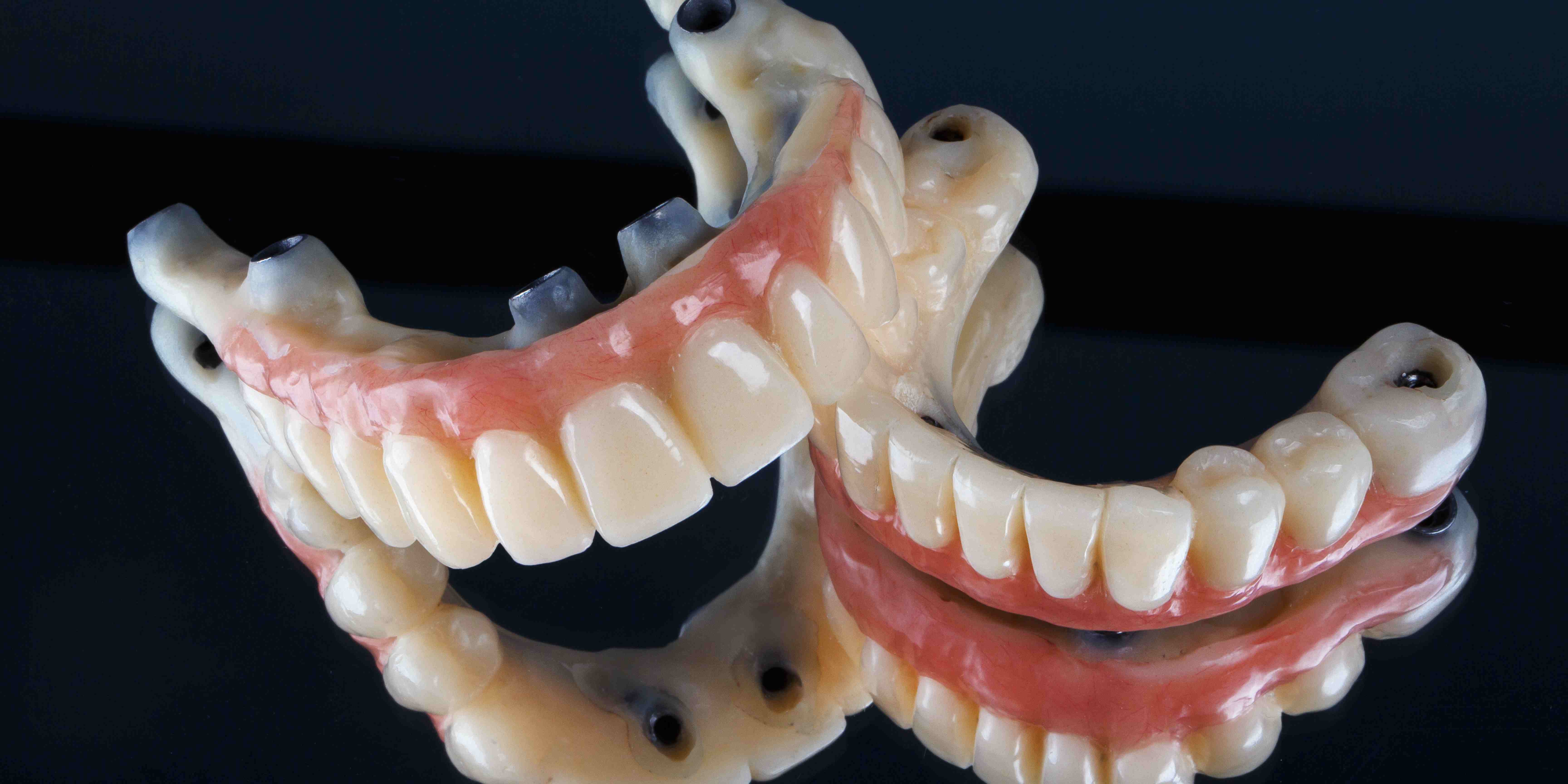
Whether you have already lost most of your teeth, struggle with loose dentures, or have teeth that can no longer be predictably restored, All-on-X treatment may help you regain a healthy, complete smile.
Read More